



New bone model could reduce reliance on animal testing in osteoporosis research
bone
Researchers at The University of Manchester and PhD students at Manchester sponsored by the Saudi Arabia government have developed a laboratory method that transforms sheep bone into a realistic model of osteoporosis, providing a potential alternative for early-stage testing of orthopaedic implants and treatments.
Researchers at The University of Manchester and Manchester Metropolitan University have developed a laboratory method that transforms sheep bone into a realistic model of osteoporosis, providing a potential alternative for early-stage testing of orthopaedic implants and treatments.
The study, published in JBMR Plus, describes how researchers used a controlled demineralisation process to alter the structure and mechanical properties of sheep bone, creating samples that closely resemble osteoporotic human bone.
Osteoporosis affects an estimated 500 million people worldwide and contributes to around 2.7 million hip fractures each year. The development of new implants and treatments requires extensive testing, often involving human cadaveric bone or animal studies, both of which can be costly, time-consuming and subject to regulatory constraints.
To address this challenge, the research team investigated whether sheep femurs sourced from the food chain could be converted into a representative model of osteoporotic bone. Sheep bone shares similarities in size with human bone and is more readily available for laboratory research.
The researchers treated sheep femurs with hydrochloric acid for different periods, removing minerals from the bone and causing progressive changes to its internal structure and strength. They then measured how the process affected bone density, architecture and mechanical performance.
“
Developing and evaluating new orthopaedic devices requires realistic models that reflect the properties of osteoporotic bone. Our findings show that a relatively simple demineralisation process can reproduce many of the structural and mechanical characteristics reported in human osteoporosis, creating a useful platform for early-stage testing and research.
Dr Glen Cooper, Reader in Medical Engineering at the Department of Mechanical and Aerospace Engineering at The University of Manchester
„

The team found that longer demineralisation times resulted in weaker, more porous bones, closely mirroring the deterioration seen in osteoporosis. Young's modulus, a measure of stiffness, fell from 110.7 MPa in untreated samples to 57.7 MPa after 96 hours of demineralisation. Volumetric bone mineral density decreased by around 33%, while porosity increased by approximately 30% compared with untreated bone.
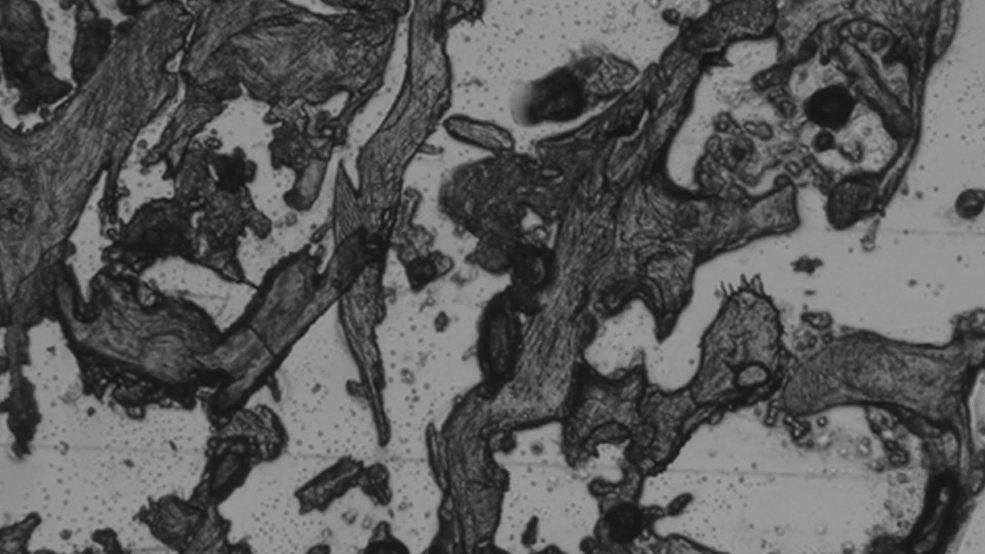
Microscopic analysis also revealed significant changes in the trabecular structure, the network of bone tissue that helps provide strength and support. As demineralisation increased, trabecular thickness decreased while trabecular separation increased, both hallmarks of osteoporotic bone.
According to the researchers, the resulting bone properties fall within ranges reported for human osteoporotic trabecular bone. This suggests the model could provide a useful and cost-effective tool for investigating osteoporosis and evaluating orthopaedic technologies before progressing to more complex testing.
The researchers note that the approach aligns with the principles of Replacement, Reduction and Refinement, often known as the 3Rs, which aim to minimise the use of animals in research where suitable alternatives are available.
The study was conducted by researchers from Manchester Metropolitan University and The University of Manchester. Lead author, Fahad Alabdah returned to Saudi Arabia as a lecturer at the University of Hail.
This research was published in: JBMR Plus
Full title of the paper: An osteoporotic bone model: developing and validating an ex-vivo bone demineralization protocol
DOI: 10.1093/jbmrpl/ziag069
URL: https://doi.org/10.1093/jbmrpl/ziag069