A new device, developed by University of Manchester PhD student Emma Biglin, will both increase accuracy markedly and reduce the numbers of animals needed to conduct radiotherapy research.
When it was discovered in 2013 that the radiotherapy doses delivered by research scientists to mice could differ by up to 40% from the dose prescribed, the research community took stock and altered ways of working to improve their accuracy. In the five years since that groundbreaking study by Desrosiers et al. (2013), methods and systems have improved markedly, but there’s still much room for improvement.
So a new device, developed by University of Manchester PhD student Emma Biglin, will both increase accuracy markedly and reduce the numbers of animals needed to conduct radiotherapy research has been warmly welcomed by many scientists. Her work was funded by the National Centre for the Replacement, Refinement and Reduction of Animals in Research (NC3Rs).
“The machines used in preclinical research are often less advanced than their clinical counterparts, and it’s much more difficult to ensure the dose given is close to the dose received because equipment is not regularly calibrated,” says Emma. “So we thought a good way around this problem would be to assess the accuracy of the equipment using materials that mimic the tissues in mice in as realistic a way as possible.”
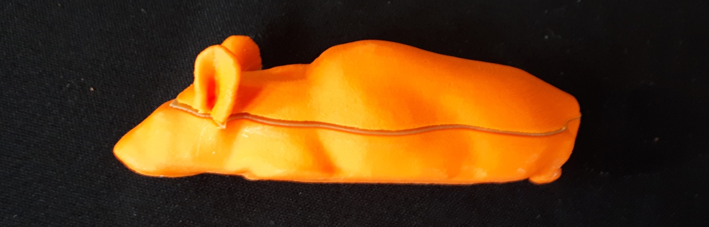
Her answer to the problem was an object shaped like a mouse made from different acrylic plastics whose materials have virtually identical densities to human bone and soft tissue. And the beauty of the creation is that it is so easy to make using 3D printing, scientists can easily design different models to perform different tasks. Once the dose received by the dummy mouse is calculated, scientists can then recalibrate the machines and the results are spectacular, with less than 5% difference between dose given and dose received; a huge improvement on previous methods.
It’s such a simple idea, but we think it will have far reaching impacts on the three Rs in radiotherapy research.
Emma Biglin / PhD student
“We know the plastics we used in our dummy mice have tissue equivalent densities – and have shown quite this clearly using CT scanning,” she says. “So that gives scientists a lot of scope to test their equipment very accurately. And the mouse shape means it is possible to identify and irradiate equivalent areas to specific organs without actually subjecting any live mouse to radiotherapy.”
The mouse dummies can be printed in sections with cavities which can house different types of radiation detectors in different parts of the dummy mouse body. For example, a piece of radiochromic film is placed in between the sections allowing researchers to see what measure the dose received by the dummy. Emma is also working with Alice Porter in the Department of Physics to develop tiny diamond detectors based on technology from CERN’s large hadron collider to enable even greater accuracy to be achieved in the future.
Reducing numbers
Because scientists have more confidence in the accuracy of their research, animal numbers can be reduced, and toxicity minimised, without affecting the statistical validity of results. Researchers will also be able to use the dummies to design experiments and finalise the set-up of the animals – a powerful refinement which will reduce discomfort and stress in the animals.
The mouse dummy is now being tested in facilities across the UK and the feedback so far, says the researcher, has been positive, though there is some way to go before they are used as standard. Anyone who has access to a 3D printer will be able to create their own mouse by accessing the files which will soon be open source.
“Making the software open source is particularly important to us, as this is all about animal welfare – as well as scientific accuracy,” says Emma. “And we’re absolutely delighted with how successful our mouse dummies have been.
“It’s such a simple idea, but we think it will have far reaching impacts on the three Rs in radiotherapy research.”